Lichens are amazing organisms. They are all around us and we hardly notice them. Found on soil, tree bark, rocks and even some under water, they are actually two organisms living together (symbiosis). The major component is a fungus (mycobiont), hence they are classified as fungi — the vast majority being ascomycetes. The other component is photosynthetic (photobiont) and may be green algae or cyanobacteria (once known as blue-green algae) or sometimes both. The photobiont can make food — sugar. The fungus can kill some of the algae cells or penetrate the algae cells to obtain food. So… the symbiotic relationship is actually a controlled parasitism. The algal cells, however, are protected from damaging excess light. Lichens are fungi that have taken up farming, and they are known as lichenized fungi.
Certainly these complex organisms can inhabit many conditions and substrates that would deter other kinds of species — hence they are known as pioneer organisms in ecological succession.
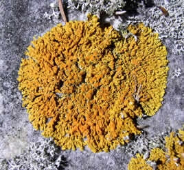
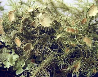
In order to help organize the lichens for identification, they are categorized by growth form of the thallus (vegetative body of the lichen). There are four major growth forms — crustose, foliose, fruticose and squamulose.
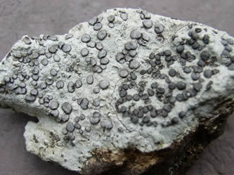
Figure 1A: Crustose lichen on rock – Smoky-eye boulder lichen, Porpidia albocaerulescens
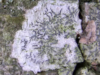
Figure 1B: Crustose lichen on bark – sexual fruiting areas are elongate (lirellae) Graphis scripta
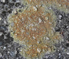
Crustose lichens (see Figures 1A, 1B, 1C) are varied, but are always firmly attached to the substrate. One must remove a portion of the substrate to remove the lichen intact. Crustose lichens have no lower layer of the thallus.
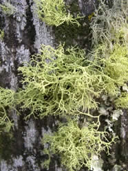
Fruticose lichens are shrubby or pendant in appearance (see Figures 3A, 3B) with no upper or lower layers.
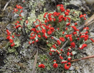
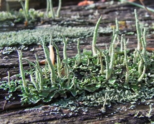
Figure 4A: British soldiers, Cladonia cristatella
Figure 4B: Common powderhorn, Cladonia cf. coniocraea
Squamulose lichens have small scale-like lobes like roof shingles. Some lichens are combinations of squamulose and fruticose such as species of Cladonia (see Figures 4A, 4B).
When a lichen reproduces sexually, it is the fungal component that makes structures called apothecia or perithecia. Apothecia look like cups or discs on the surface of the thallus. Perithecia are flask shaped structures mostly embedded in the thallus. Within these sexual structures you will find asci containing the fungal spores. When released, spores must find algal cells in order to lichenize. The spore structure is important in identifying the crustose lichens. Sometimes the crustose lichens are called microlichens because you do need a compound microscope for proper ID. Field guides to the macrolichens (foliose and fruticose) usually identify the species by substrate first, then by growth form followed by color. Another feature that is critical to observe with a 10x loupe relates to how the lichens reproduce asexually by vegetative means (the major reproductive strategy). Soredia are vegetative propagules containing algal cells and fungal hyphae. They form in cracks of the thallus (soralia), and can be dispersed by wind, insects, birds and water. With magnification, soredia can look powdery or like tiny crystals. Other propagules look like upright fingers or coral projections, and represent growths of the top layer (upper cortex) with algae underneath. These projections which are called isidia can break off and disperse. Chemical spot tests are also used in identification: K = 10% KOH, C = bleach, PD = para-phenylenediamine. In addition, TLC (thin-layer chromatography) and observing the reaction to long wave UV light can be useful for ID (see Brodo, Lichens of North America, pages 103-110).
Some of the most fascinating facts on lichens (listed below) relate to their connections to the ecosystem and how humans have used them in history and currently.
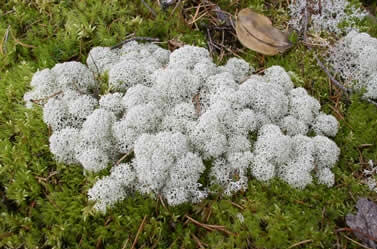
Figure 5: One of the reindeer lichens, Cladonia stellaris
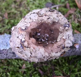
Figure 6: Nest of a Ruby-throated hummingbird
Eco-Connections
many lichens are air pollution indicators (sensitivity to SO2, etc.)
lichens containing cyanobacteria can fix nitrogen
8% of the earth’s terrestrial surface is dominated by lichens
90% of the winter diet of caribou (“reindeer moss” are lichens) (Figure 5)
food for mule deer, mountain goats, pronghorn antelope, northern flying squirrels, western voles, wild turkey, slugs, snails, mites, springtails, certain caterpillars
nesting material for 50 species of birds (Figure 6)
background camouflage for lizards, certain moths, tree frogs; lacewing larvae cover wings with lichens
sand and soil stabilizers (microbiotic crusts)
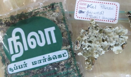
Figure 7: Kal Paasi spice
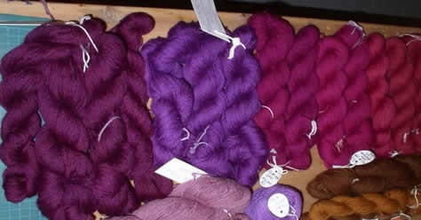
Figure 8: Purple dyed wool from Ochrolechia sp. – photo taken at the International Fungi Fiber Symposium in Denmark, 2005
Human Uses
food for the Salish (west coast tribe)
Japanese delicacy – Umbilicaria esculenta
food in times of famine – Cetraria islandica ground – up and added to flour
spice used in India – Kal Paasi (Figure 7)
clothing – west coast pendant lichens woven into moccasins, leggings
royal purple dye from Ochrolechia (cudbear) (Figure 8) and Roccella (orchil)
litmus pigment
brown pigments for Harris tweeds and Navajo rugs
perfume fixatives from Evernia prunasti
antibiotic properties of Usnea , Cetraria
wolf lichen, Letharia vulpina, added to bait to poison wolves
décor: model train landscaping and flower arranging- Cladonia spp.
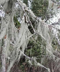
Figure 9A: Lace lichen – Ramalina menziesii
Figure 9B: Lace lichen – Ramalina menziesii
Get to know the lichens. They are fascinating organisms. You can study them at any time of the year and in any weather conditions. Congratulations to California. In 2016, they became the first state to have a “State Lichen” Ramalina menziesii,the Lace Lichen (Figures 9A and 9B).
REFERENCES
Brodo, Sharnoff and Sharnoff. 2001. Lichens of North America. Yale Univ. Press.
Hinds and Hinds. 2007. Macrolichens of New England. NY Botanical Garden Press.
McMullin and Anderson. 2014. Common Lichens of NE North America. NY Botanical Garden
Nash, Thomas. 2008. Lichen Biology. Cambridge University Press.
Purvis, William. 2000. Lichens. Natural History Museum.
Walewski, Joe. 2007. Lichens of the North Woods. North Woods Naturalist.
NAMA Store >